2.1. Linear movements
All the voluntary and involuntary muscular movements of our body derive from natural nanomotors that develop linear movements. Very complex protein molecules called myosins are responsible for these movements. Such molecules consist of long tails to which two large heads are connected (Fig. 10a, top right).
In muscle cells (Fig. 10a), many myosin molecules are assembled through the tails to give a filament from which the heads extend; these attach themselves to other filaments, called actin, parallel to those of myosin and acting as a guide. A chemical reaction involving the adenosine triphosphate species (ATP, Fig. 11) provides energy to the system that is used to radically change the shape of the myosin heads and to force it, as a consequence of this change of shape, to move along the actin filament (Fig. 10b).
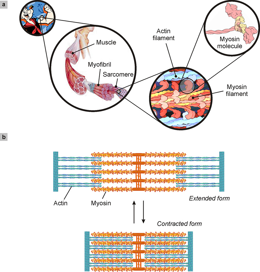
Fig. 10. Muscle function. (a) Schematic representation of the myosin molecule which has a long tail to which two large heads are attached. In muscle cells, many myosin molecules are intertwined by the tails to give a filament from which the heads extend. These attach themselves to other parallel filaments consisting of molecules of another protein, actin. These filaments, which are able to glide over one another, are contained in sarcomeres. (b) Muscle contraction is generated by the chemical reaction described in Fig. 11 which modifies the shape of the heads of the myosin molecules forcing them to glide over the actin filaments. The sarcomere thus passes from an expanded form to a contracted form. Credits: RCSC PDB and David S. Goodsell, The Scripps Research Institute, La Jolla, USA. Here, an explanatory movie: The Inner Life of the Cell. Courtesy BioVision, Harvard University. Copyright © 2006 The President and Fellows of Harvard College.
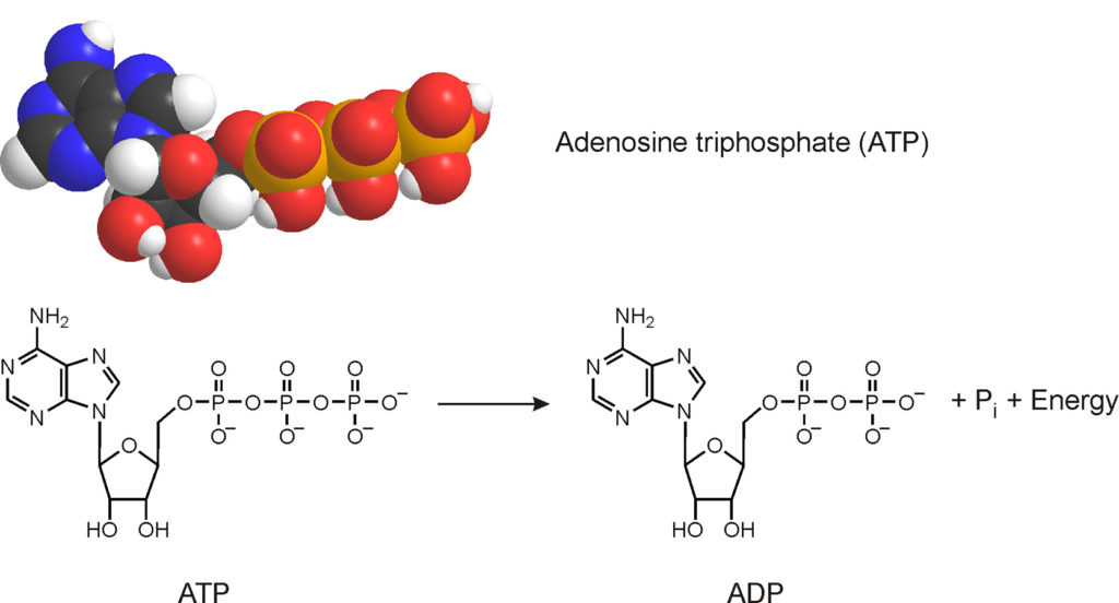
Fig. 11. The adenosine triphosphate (ATP) molecule is the energy currency of cells. By breaking the ATP molecule to give adenosine diphosphate (ADP) and phosphate (Pi) energy is obtained, used to feed cellular functions.
In a rapidly contracting muscle, each unit of myosin moves five times per second, covering a distance of about 10 nm with each movement. It is estimated that two billion nanometric movements are needed to generate the force required to catch a baseball.
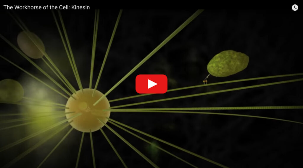
The task of linear molecular motors is not limited to muscle contraction. A real railway network operates inside the cells to transport substances from one side of the cell to the other. These nanometric “freight trains” are driven by linear molecular motors such as kinesin and dynein.
Kinesin, for example, consists of a motor part, possessing two heads, and an area intended for the capture and release of the cargo to be transported (Fig. 12). The two heads of the motor part, due to chemical reactions involving ATP that they can accommodate, alternatively bind and dissociate from a microtubule (a protein filament 30 nm wide and 50000 nm long), practically walking on it in 72 nm steps, at a speed of 1000 nm per second.
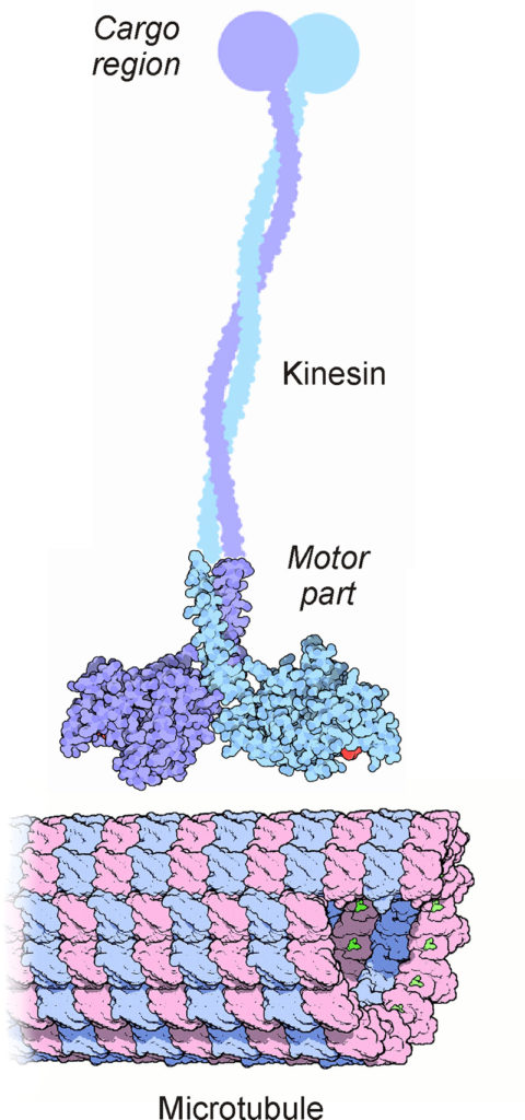
Fig. 12. Kinesin is a linear molecular motor capable of carrying a load along a track consisting of a microtubule. Also in this case the energy required to operate the motor part of the kinesin comes from the breakup of ATP molecules in particular sites located in the heads. It should be noted that the various parts of the figure are not to scale: the microtubule is much larger than the kinesin. Credits: RCSC PDB and David S. Goodsell, The Scripps Research Institute, La Jolla, USA. Here, an explanatory movie: The molecular mechanism of muscle contraction (Copyright reserved) and another one below: